When a parent notices a persistent lump in their child's neck, it can understandably cause significant concern. Among the various causes of pediatric neck masses, a branchial cleft cyst (BCC) is a relatively common congenital anomaly.
These cysts arise from remnants of the embryonic branchial apparatus—the structures that form the neck and throat during fetal development. When these structures don't fully disappear as intended, they can form a fluid-filled sac (a cyst) that may manifest later in childhood.
Managed by the Department of Paediatric Surgery at St. Stephen’s Hospital, New Delhi
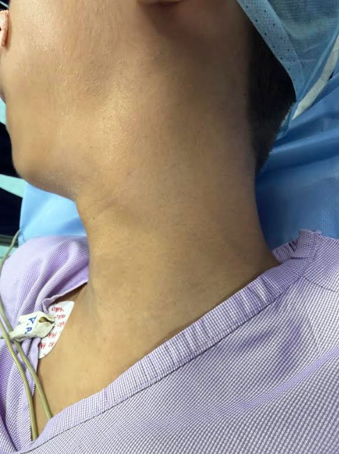
To better illustrate how these are managed in a clinical setting, we are sharing a recent case involving a 12-year-old boy. He presented to our clinic with a persistent, painless swelling on the left side of his neck that had been present for three months.
The mass was noted to be gradually increasing in size. Importantly, the patient reported no associated symptoms such as fever, pain, or difficulty swallowing (dysphagia), which is typical for non-infected branchial cleft cysts.
Upon clinical examination, we observed:
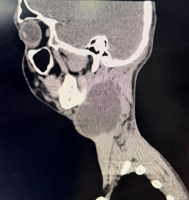
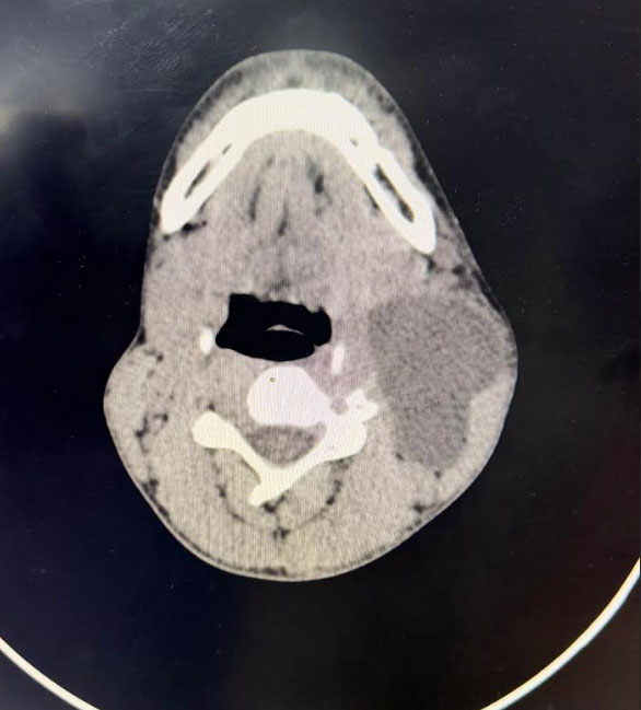
To evaluate the extent of the mass and plan the surgical approach, a CT scan of the neck was performed. The imaging provided clear, actionable data:
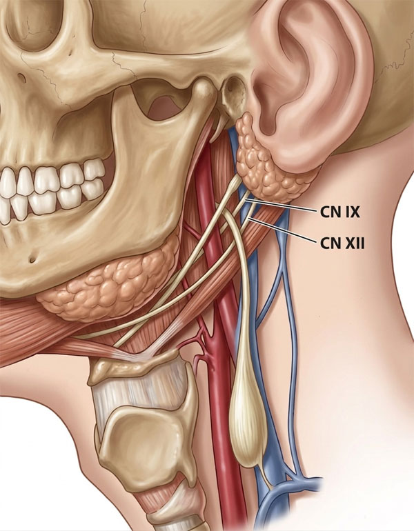
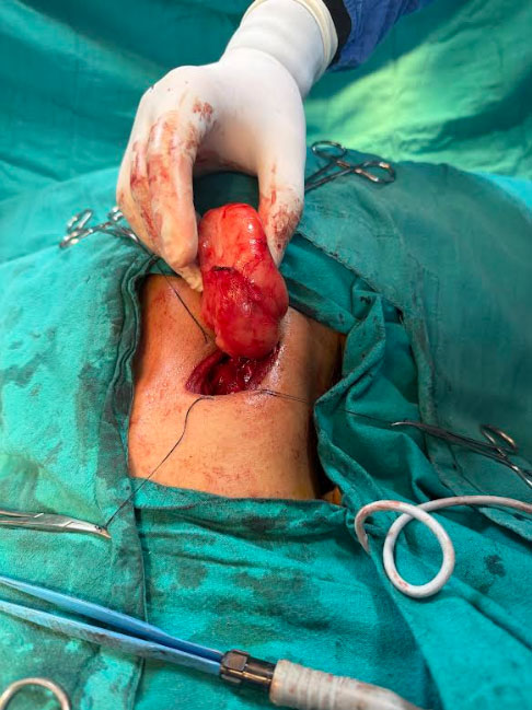
Given the size of the cyst and the proximity to vital structures, surgical intervention was necessary. The patient underwent a formal excision of the mass. The surgical objective was twofold: ensure the complete removal of the cyst wall to prevent recurrence, while meticulously preserving the adjacent neurovascular bundle – Mainly the Carotid Artery and the IX and XII Cranial nerves.
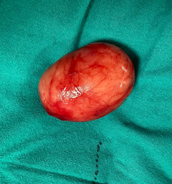
We are pleased to report that the surgery was successful, and the mass was excised in its entirety.
The final histopathology report definitively confirmed the diagnosis: Branchial Cleft Cyst.
At his most recent follow-up appointment, the patient remains completely asymptomatic and is recovering excellently. This case serves as a powerful reminder of the vital role that thorough diagnostic imaging and surgical precision play in the successful management of pediatric neck masses.
While many neck masses in children are benign, any persistent, enlarging, or symptomatic lump should be evaluated by a Paediatric Surgeon. Key signs to watch for include:
Early assessment ensures an accurate diagnosis and timely treatment, providing the best possible outcome for the child.